Neuromodulation

Neuromodulation

Please download the pamphlet for more information on neuromodulation for pain, spasticity and other neurologic disorders.

Neuromodulation Pamphlet

In general, neuromodulation involves interfacing the nervous system with circuits or implanting catheters to deliver medication directly. This allows for direct treatment of a variety of neurologic problems including pain, spasticity from spinal cord injury, brain injury or multiple sclerosis, movement disorders and others.

Deep Brain Stimulation

Deep Brain Stimulation

What is Deep Brain Stimulation (DBS)?

DBS procedure involves the implantation of electrode arrays into the brain and the insertion of the implantable pulse generators (IPGs) that drive the system. DBS alleviates various symptoms of many neurological disorders by applying electrical fields in specific regions of the brain. Currently, DBS is being used predominantly for people suffering from movement disorders such as Parkinson’s disease and essential tremor. But other indications are on the horizon. Unlike other forms of movement disorder surgery (i.e. pallidotomy and thalamotomy), DBS can be modified over time and is fully reversible.

The operation is done in two stages. The first stage requires a postoperative, overnight stay. The DBS leads themselves are implanted stereotactically, using computer aided targeting while the patient is awake. We use microelectrode recordings and microstimulation as well as macrostimulation to accurately guide and position the electrode array. This stage of the implantation is all done through a 14mm (less than half an inch) hole. The second stage is an out-patient procedure. The IPG is connected to the DBS lead. Under general anesthesia this connection is tunneled under the skin to a pocket under the collar bone, where the battery pack and pulse generating unit are placed. This procedure takes about 45 minutes for each IPG. After recovery the patient is sent home with wound care instructions and a DBS patient information booklet. Follow-up with the neurologist and neurosurgeon with adjustments to the IPG and wound checks are scheduled before the patient leaves the hospital. IPG adjustments are done in the office using a magnet over the unit without even removing any clothing. These adjustments are done in conjunction with medication adjustments.

DBS Post-operative Guide

Our Philosophy

At National Capital Neurosurgery we emphasize a team approach to diagnosis, management, surgical targeting and follow-up care for DBS. Our team includes a neurosurgeon, neurologist, and neurophysiologists, who all have experience in movement disorders and their management. We believe this team approach provides the patient the best possible care. Consultation with both the neurosurgeon and the neurologist are necessary prior to any decisions about eligibility for surgery. The neurosurgeon and neurophysiologist work together in the operating room to ensure accurate targeting and the best results possible from the surgery. Follow-up care is done both by the neurologist and the neurosurgeon. The neurologist manages the actual programming of the stimulator and the neurosurgeon follows the patient for wound healing and other surgical issues. The patient’s care and progress is the essential focus for the team.

Who are Eligible Candidates?

Individuals suffering from Parkinson’s disease (PD) who are not tolerating medical therapy or who cannot tolerate some of the side effects of the medications (dyskinesia in particular) are eligible candidates for DBS. Medical therapy is the first line of treatment in PD and thorough trial of medications and extensive follow-up with a neurologist is important before surgery can be considered. Those individuals who have benefited from or responded well to levodopa or Sinemet therapy are better candidates. Previous pallidotomy or thalamotomy does not exclude one from DBS. In fact patients who have benefited from these procedures will most likely benefit from DBS. All cardinal features of PD are treated with DBS. Freezing, stiffness, and tremor all show improvement with DBS in the subthalamic nucleus. Dyskinesia also shows improvement as the amount of medication is decreased as the stimulator is adjusted. Those with essential tremor (ET) who are growing “resistant” to the medication or who are suffering from medication side effects, are good candidates for thalamic DBS. Extensive work-up and medical therapy should always precede surgery for ET.

How Do I Get Started?

If you wish to have an initial consultation, you may call (301) 718-9611 and ask for a DBS consultation with Dr. Zachary Levine. Please bring with you a list of medications and your dose schedule. We will need this for our records. In addition, we will need the name of your primary care physician as well as your treating neurologist. If you have had any MRI or CT scans of your brain, please bring them or arrange to have copies sent to our office.

Cranial Surgery

Cranial Surgery

Cranial surgery includes surgery for tumors, vascular abnormalities (aneurysms, arteriovenous malformations, etc), blood clots, seizures, hydrocephalus, and Deep Brain Stimulation for neurologic diseases. Neurosurgeons work with other specialists in and outside of the operating room to get the best results possible. We often use minimally invasive or noninvasive techniques. Image guided, computer assisted and endoscopic surgery is also done in the brain.

Brain Tumors

Brain tumors include benign and more aggressive tumors, both of which often need surgical treatment or biopsy for diagnosis. Surgery for brain tumors ranges from minimally invasive biopsies or nearly non-invasive radiosurgery, to more aggressive surgical approaches. Some tumors require more than one modality for diagnosis and treatment.

Surgery for brain tumors is often done for resection or removal of accessible lesions. Modern techniques such as image-guided surgery, contact lasers and endoscopes have made the treatment of these tumors easier on the patient. Many types of brain tumors can be addressed surgically, including meningiomas, schwannomas, gliomas, metastatic tumors, pituitary adenomas, and others. Dr. Levine has published on the surgical treatment of meningiomas and has taught courses on resection of these tumors at the cranial base.

Open surgery also allows the use of other treatment during surgery. Chemotherapy and radiation can be delivered directly to the tumor during an open operation. This allows for higher concentrations of drug or radiation to be delivered to the site without exposing the rest of the body. Tumors at the base of the skull often require more extensive procedures and more lengthy operations. These tumors often involve the nerves and vessels that exit and enter the skull. Special procedures and monitoring are used during the surgery to increase the safety of the patient.

Deep Brain Stimulation

Deep Brain Stimulation (DBS) is used to treat movement disorders such as Parkinson’s Disease, Essential Tremor and Dystonia. Other disorders that are under investigation for treatment include: Tourette’s Syndrome, Obsessive Compulsive Disorder and Depression. Dr. Levine has extensive experience in DBS and has taught DBS nationally and internationally. He is a Key Opinion Leader in DBS. He has innovated the field with the propagation of frameless techniques for DBS and collaborates with the NIH.

Epilepsy Surgery

Epilepsy is most commonly treated with medication. However, in some circumstances surgery is needed to eliminate the focus of seizures or a device may be implanted to decrease the number of seizures. Dr. Levine works with epileptologists and the epilepsy monitoring unit at Holy Cross Hospital to help screen and eventually treat surgically those patients that require this approach.

Hydrocephalus

Is often associated with headaches, gait disturbance, nausea and difficulty with thinking. It is associated with abnormal flow of the cerebrospinal fluid. There are a number of procedures that can be used to treat this condition. Often a shunt is used to divert the fluid to the abdomen or heart. In some cases an endoscopic brain surgery can be used to treat the condition as well. This is called an endoscopic third ventriculostomy. Dr. Levine has experience with both forms of treatment and can determine which is appropriate with the history and imaging studies provided at consultation.

Radiosurgery

Radiosurgery is a technique used to treat tumors or vascular malformations of the brain without cutting the skin. Carefully designed radiation beams are delivered to the site, minimizing damage to surrounding structures. This is ideal for patients with multiple tumors of the brain. Radiosurgery is also used when conventional surgery might be too dangerous. Radiosurgery is not useful for every situation but plays a very important role in the surgical treatment of brain tumors. Stereotactic radiosurgery is a minimally invasive alternative to conventional brain surgery in some circumstances. This procedure allows safe and effective treatment for many types of intracranial tumors and vascular malformations. By using radiation sources with the aid of sophisticated dose planning software, radiosurgery can often treat conditions once considered inoperable. A linear accelerator delivers beams or X-rays that converge on and destroy tumor cells and abnormal blood vessels, such as arteriovenous malformations (AVMs).

BrainPath

BrainPath is a new and unique way to access deep areas of the brain using a minimally invasive technique. This allows the resection of deep seated tumors and evacuation of blood clots deep in the brain that were traditionally considered inaccessible. Dr. Levine is currently the first surgeon to do this procedure in the metropolitan DC area. Holy Cross Hospital has the first fully functional BrainPath center in the state of Maryland and the DC area! Dr. Levine is willing to consult on patients who may be candidates for this technique.

Cerebrovascular Disease

Blood flow to brain can be interrupted causing a stroke. This can result in a blood clot in the brain that might need to be removed. Also some strokes cause intense swelling in the brain and surgical decompression can be used to lower the pressure in the skull. Other patients have vascular abnormalities in the brain such as aneurysms, or arteriovenous malformations. These can often be treated with endovascular techniques. These too are serious and often require emergency surgery for treatment.

Spinal Surgery

Spinal Surgery

Disorder of the Spinal Column

Spine surgery includes treatment of the spinal column, or the support structures around the spinal cord and spinal nerves. Compression or constriction of the nerves and spinal cord by the bony overgrowth of the spine or herniated disks can cause pain, sensory changes or weakness. Treatment often requires decompression (removal of abnormal bone growth/herniated disks) of the spinal cord and nerves to alleviate symptoms. Spinal column disorders requiring surgery range from degenerative processes to traumatic injury to tumors. In some circumstances, the stability of the spine is affected. When needed, spinal fusion with or without instrumentation or “hardware” is used to re-establish spinal stability. Minimally invasive techniques and microsurgical techniques are utilized when appropriate to get the best results for the patient.

SpinalCordNCN

Regions of the Spine

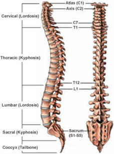
Humans are born with 33 separate vertebrae. By adulthood, we typically have 24 due to the fusion of the vertebrae in the sacrum.

  • The top 7 vertebrae that form the neck are called the cervical spine and are labeled C1-C7. The seven vertebrae of the cervical spine are responsible for the normal function and mobility of the neck. They also protect the spinal cord, nerves, and arteries that extend from the brain to the rest of the body.
  • The upper back, or thoracic spine, has 12 vertebrae, labeled T1-T12.
  • The lower back, or lumbar spine, has 5 vertebrae, labeled L1-L5.
  • The lumbar spine bears the most weight relative to other regions of the spine, which makes it a common source of back pain.
  • The sacrum (S1) and coccyx (tailbone) are made up of 9 vertebrae that are fused together to form a solid, bony unit.

Disorder of the Spinal Cord

Spinal cord disorders that require surgery include tumors or structural abnormalities of the spinal cord. Spinal cord tumors range from benign to aggressive and surgery or biopsy might be needed to obtain a diagnosis and aid in treatment. Some tumors can be completely treated with surgery alone, others might require other therapies in conjunction with surgery. Structural abnormalities of the spinal cord can include dilated, fluid-filled channels in the spinal cord (syrinx, syringomyelia), tethered spinal cords and other developmental problems. Although most of these present in childhood, some become problems as we get older. Surgery in these circumstances helps to alleviate symptoms by altering the structure of the spinal cord.

Disorder of the Spinal Nerve

The problems involving the spinal nerves often come from compression. Degeneration and herniation of the disks in the neck, back and low back can compress spinal nerves as they exit the spinal column. Symptoms can range from numbness to pain to weakness in an arm or leg and difficulty walking. Surgery might include removal of all or part of the disk. If the whole disk is removed, spinal fusion is usually required to maintain the stability of the spine. Degeneration of joints in the spine can also cause spinal nerve compression giving very similar symptoms. Finally, tumors of the nerves themselves can cause pain and dysfunction. All of these can be treated surgically if indicated.

Tumors of the Spine and Spinal Cord

Less common than disorders of the structural elements of the spine, spinal tumors and tumors of the spinal cord also treated by neurosurgeons. These tumors can be metastatic from other parts of the body or can spread from tumors near the spine. These are often cancers and will require other forms of treatment along with or instead of surgery. The tumors of the spinal cord and the nerve roots themselves are often benign and can be treated with surgery alone. We use microsurgical techniques and contact lasers and electrophysiologic monitoring of the spinal cord to improve resection and increase safety.

Surgical Procedures

Most of the surgical procedures for the spine are for decompression of the spinal nerves or spinal cord. The compression can cause pain, weakness and sensory changes that can be reversible with decompression. Surgery is not always the first choice for treatment, but it may be necessary for certain circumstances. Minimally invasive approaches are used, when possible to minimize postoperative discomfort and hasten recovery. The acronym MIS indicates Minimally Invasive Surgery. Listed below are some of the more common procedures performed. This is not a comprehensive list but describes the most common operations done.

  • MIS Microdiscectomy

Using a tubular retractor system through a 2cm (less than 1 inch) incision we are able to remove a herniated disk and decompress nerve roots. Using an operating microscope and microsurgical instruments increases visualization and decreases the “footprint” of the operation. Intra-operative imaging increases the accuracy of the surgery. The surgery takes about 45 minutes and most patients go home the same day. This one of the most common spinal surgeries performed.

  • Spinal Stabilization

Often decompression of the nerve roots is coupled with a stabilization procedure. This is not a fusion. This procedure allows for decompression of the nerve roots and stabilization of the spine without a bony fusion. Spinal stabilization is done in cases of misalignment of the vertebrae without instability. A decompression might result in instability. After the decompression is done a stabilization device is applied to the spinal segment to prevent instability and preserve motion of the spine. The device used at National Capital Neurosurgery is the coflex. This is often done as an outpatient procedure.

  • Lumbar Fusion (Open and MIS)

In cases of instability of the spine associated with spondylolisthesis, a lumbar fusion with decompression is needed. This can be done using an MIS technique using percutaneous pedicle screws or cortical bone screws in an open technique. Fusion is used in cases of instability or fracture. This operation decompresses the spine and fuses the segment(s) that are unstable.

  • Cervical Fusion

When cervical disks degenerate this can result in nerve root or spinal cord compression. When surgery is needed this often requires complete discectomy and fusion. This is done from an incision in the front of the neck. Microsurgical techniques use sophisticated neurophysiologic monitoring to increase the safety of the surgery. Cervical fusion is often done as an outpatient procedure.

  • Cervical Arthroplasty

When cervical disk herniations present with otherwise normal anatomy and no significant degeneration of the joints, an artificial disk can be used instead of fusion. Unlike a cervical fusion, the arthroplasty maintains normal motion. It is done using the same microsurgical technique as a fusion, but an artificial disk is used instead of a fusion device. This, too, is almost always an outpatient procedure.

  • Deformity and Scoliosis

Often abnormal curvature of the spine can lead to pain and neurologic compromise. This can be due to congenital issues, degeneration or from previous surgery. In some circumstances, this “deformity” needs to be corrected. This requires expertise and judgment. Sometimes minimally invasive techniques can be used to achieve the result with short hospital stays and less postoperative discomfort.